Data information
To register, please download our registration form and send to the organizers.
We provide 194 LGE MRIs. All these clinical data have got institutional ethic approval and have been anonymized (please follow the data usage agreement, i.e., CC BY NC ND). The provided gold standard labels include: left atrial (LA) blood pool (atriumSegImgMO.nii.gz) and LA scars (scarSegImgM.nii.gz).

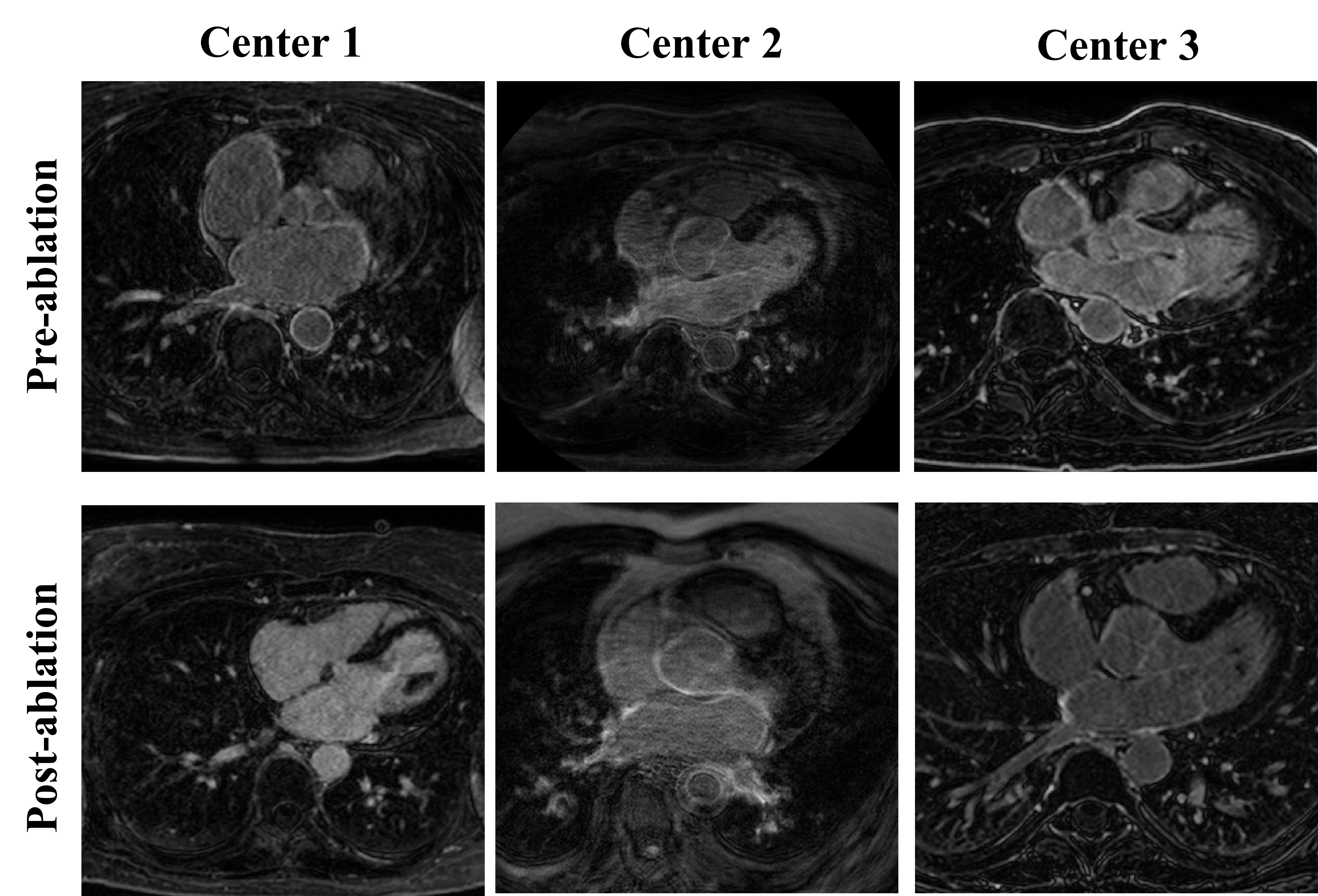
The details of LGE MRIs are as follows:
Center 1 (University of Utah): The clinical images were acquired with Siemens Avanto 1.5T or Vario 3T using free-breathing (FB) with navigator-gating. The spatial resolution of one 3D LGE MRI scan was 0.625 × 0.625 × 2.5 mm. The patient underwented an MR examination prior to ablation or was 3-6 months after ablation.
Center 2 (Beth Israel Deaconess Medical Center): The clinical images were acquired with Philips Acheiva 1.5T using FB and navigator-gating with fat suppression. The spatial resolution of one 3D LGE MRI scan was 1.4 × 1.4 × 1.4 mm. The patient underwented an MR examination prior to ablation or was 1 month after ablation.
Center 3 (King’s College London): The clinical images were also acquired with Philips Acheiva 1.5T using FB and navigator-gating with fat suppression. The spatial resolution of one 3D LGE MRI scan was 1.3 × 1.3 × 4.0 mm. The patient underwented an MR examination prior to ablation or was 3-6 months after ablation.
Note that the resolution of images in validation/test dataset were adjusted to 1.0 × 1.0 × 1.0 mm for convenience.[2] Lei Li, Veronika A Zimmer, Julia A Schnabel, Xiahai Zhuang*: Medical Image Analysis on Left Atrial LGE MRI for Atrial Fibrillation Studies: A Review, Medical Image Analysis, vol. 77, 102360, 2022. link.
[3] Lei Li, Veronika A Zimmer, Julia A Schnabel, Xiahai Zhuang*: AtrialGeneral: Domain Generalization for Left Atrial Segmentation of Multi-Center LGE MRIs, MICCAI, 557–566, 2021. link