The gold standard segmentation of test data have been released!!!
The event is closed, but the MSCMR data with gold standard are available after registration.

For participants who want to download and use the data, they need to agree with the conditions above and the terms in the registration form (please sign the form and send to the organizers.)
Agree and download

The Recipient(s) of these data commit to not disseminate the data to any third party. Please cite these two papers when you use the data for publications.
[1] S Gao, H Zhou, Y Gao, X Zhuang. BayeSeg: Bayesian Modeling for Medical Image Segmentation with Interpretable Generalizability. Medical Image Analysis 89, 102889, 2023 code&tutorial, link (Elsevier-MedIA 1st Prize & Best Paper Award of MICCAl society 2023)
[2] Xiahai Zhuang: Multivariate mixture model for myocardial segmentation combining multi-source images. IEEE Transactions on Pattern Analysis and Machine Intelligence (T PAMI), vol. 41, no. 12, 2933-2946, 2019. link code
[3] F Wu & X Zhuang. Minimizing Estimated Risks on Unlabeled Data: A New Formulation for Semi-Supervised Medical Image Segmentation. IEEE Transactions on Pattern Analysis and Machine Intelligence (T PAMI) 45(5): 6021 - 6036, 2023 link code

Cardiac cross modality domain adaptation works from zmic link
Wu,FP's medical cross modality domain adaptation work & Data link-CO LGE CMR patchs

Clinical Background
Accurate computing, analysis and modeling of the ventricles and myocardium from medical images is important, especially in the diagnosis and treatment management for patients suffering from myocardial infarction (MI).
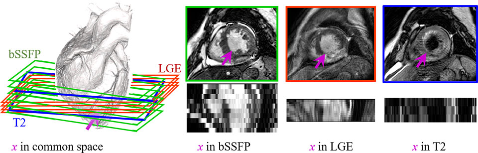
MRI is particularly used to provide imaging anatomical and functional information of heart, such as the T2-weighted CMR which images the acute injury and ischemic regions, and the balanced-Steady State Free Precession (bSSFP) cine sequence which captures cardiac motions and presents clear boundaries. Particularly, LGE CMR can enhance the infarcted myocardium, appearing with distinctive brightness compared with the healthy tissues. It is widely used to study the presence, location, and extent of MI in clinical studies. Thus, delineating ventricles and myocardium from LGE CMR images is important.
However, the segmentation is still arduous, particularly due to the pathological myocardium from LGE CMR; but manual delineation is generally time-consuming, tedious and subject to inter- and intra-observer variations.
Motivation
This challenge aims at creating an open and fair competition for various research groups to test and validate their methods, particularly for the multi-sequence ventricle and myocardium segmentation.
We provide 45 multi-sequence CMR images from patients who underwent cardiomyopathy. Each patient had been scanned using the three CMR sequences, i.e. the LGE, T2 and bSSFP. The task of this challenge is to segment the ventricles and myocardium from LGE CMR, combing with other two sequences (T2 and bSSFP) from same patients, which can be used to assist the LGE CMR segmentation. This challenge is not only to benchmark various segmentation algorithms, but also to cover the topic of general cardiac image segmentation, registration and modeling.
Important date
Abstract registration: July 1st, 2019 new
Test results submission: July 7th, 2019 extended from July 1st, 2019
Test Performance Feedback: July 10th, 2019 new
Paper submission: July 16th, 2019 new
Notification of acceptance: August 6th, 2019 extended from August 1stextended from July 3rd, 2019
Workshop Camera ready: August 15th, 2019 extended from July 17th, 2019
Topic
The segmentation of ventricles and myocardium from LGE CMR:
Segmentation strategies
Direct segmentation on the LGE CMR dataset
Segmentation with the assistance from the other two
      CMR sequences from the same patients
Others
Segmentation algorithms
Atlas based segmentation
Statistical shape model based segmentation
Deep learning segmentation
Others
Award
The best works will be awarded with prizes, and the selected papers will be published in Lecture Notes in Computer Science (LNCS), Springer with STACOM workshop (see previous proceedings).
Note: Paper, presentation and test results will all be taken into account when a piece of work is evaluated.
Result: