Data information
For participants who want to download and use the data, they need to agree with the conditions above and the terms in the registration form (please sign the form and send to the organizers.)
Agree and download from FDU server or
Agree and download from mega link.
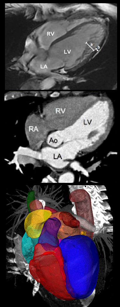
We provide 120 multi-modality whole heart images from multiple sites, including 60 cardiac CT/CTA and 60 cardiac MRI in 3D that cover the whole heart substructures. All these clinical data have got institutional ethic approval and have been anonymized.
The data were collected based on in vivo clinical environment and the data were used in clinics. So the data had various image quality, some were with relative poor quality. However, it is necessary to include these datasets to validate the robustness of the developed algorithms when it comes to real clinical usage.
The cardiac CT/CTA data were acquired using routine cardiac CT angiography. All the data cover the whole heart from the upper abdominal to the aortic arch. The slices were acquired in the axial view. The inplane resolution is about 0.78 × 0.78 mm and the average slice thickness is 1.60 mm. The MRI data were acquired using 3D balanced steady state free precession (b-SSFP) sequences, with about 2 mm acquisition resolution at each direction and reconstructed (resampled) into about 1 mm.
We split the datasets into training (20 CT and 20 MRI representative) and test (40 CT and 40 MRI) data sets. For the training data sets, we provide manual segmentation of the seven whole heart substructures,
(1) the left ventricle blood cavity (label value 500);
(2) the right ventricle blood cavity (label value 600);
(3) the left atrium blood cavity (label value 420);
(4) the right atrium blood cavity (label value 550);
(5) the myocardium of the left ventricle (label value 205);
(6) the ascending aorta (label value 820), which is defined as the aortic trunk from the aortic valve to the superior level of the atria; for example, the mean length of this trunk is about 41.9 mm as we measured in 25 healthy subjects;
(7) the pulmonary artery (label value 850), which is defined as the beginning trunk between the pulmonary valve and the bifurcation point, for example, the mean length of this trunk is about 41.9 mm as we measured in 25 healthy subjects.
The great vessels of interests include the ascending aorta and the pulmonary artery. Due to the different fields of view, the coverage of the great vessels can be different in different scans. To have a consistent definition across different subjects, the ascending aorta and pulmonary artery are thus defined as above, while the validation of whole heart segmentation can solely focus on the major trunk of the vessels. The manual segmentation for both training and test data generally covers beyond the defined length of the vessels. In the evaluation, we will use a method to cut the results of the vessel segmentations to the limited length as described above. For the submission, we encourage the participants to submit their segmentation of vessels as longer as possible.
[1] Xiahai Zhuang: Multivariate mixture model for myocardial segmentation combining multi-source images. IEEE Transactions on Pattern Analysis and Machine Intelligence 41(12): 2933-2946, 2019. link code[2] X Zhuang & J Shen: Multi-scale patch and multi-modality atlases for whole heart segmentation of MRI, Medical Image Analysis 31: 77-87, 2016 (link)
[3] F Wu & X Zhuang. Minimizing Estimated Risks on Unlabeled Data: A New Formulation for Semi-Supervised Medical Image Segmentation. IEEE Transactions on Pattern Analysis and Machine Intelligence (T PAMI) 45(5): 6021 - 6036, 2023 link code
[4] S Gao, H Zhou, Y Gao, X Zhuang. BayeSeg: Bayesian Modeling for Medical Image Segmentation with Interpretable Generalizability. Medical Image Analysis 89, 102889, 2023 code&tutorial, link (Elsevier-MedIA 1st Prize & Best Paper Award of MICCAl society 2023)